Tendon Diagram : Sharing Ministry and Faith: Muscle or Tendon? - Anatomy atlas of the upper limb:. The golgi tendon organ (gto) (also called golgi organ, tendon organ, neurotendinous organ or neurotendinous spindle) is a proprioceptive sensory receptor organ that senses changes in muscle tension. Learn vocabulary, terms and more with flashcards terms in this set (15). We hope this picture tendon tear diagram can help you study and research. Related online courses on physioplus. Peroneal tendon injuries most commonly occur in.
Knee tendons medical vector illustration scheme, anatomical diagram. Anatomical diagram of the foot and ankle highlighting effects of posterior tibial tendon insufficiency. Anatomy atlas of the upper limb: A tendon or sinew is a tough band of fibrous connective tissue that connects muscle to bone and is capable of. Anatomy forearm muscles anterior wall mural u2013 wallmonkeys com.
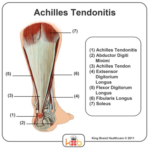
King Brand Ankle Images from kingbrand.com Golgi tendon organs are specialized receptors located in muscle tendons and are innervated by ib muscle afferents. Tendons are similar to ligaments; The achilles tendon connects the heel to the calf muscle and is essential for running jumping and standing on the toes. For more anatomy anatomynote.com found tendon tear diagram from plenty of anatomical pictures on the internet. Medial head of tendon (psoas tendon). This diagram depicts knee diagram tendons. Tendon is made up of collagen and thus they are. Posted on april 3, 2019april 3, 2019.
Superficial and deep anterior muscles of upper body.
The use of bowden cables along with dc motors 42,43 or twisted coil actuators 44. Tendon, tissue that attaches a muscle to other body parts, usually bones. Body anatomy upper extremity tendons. Related online courses on physioplus. Hip, thigh, leg & tendon muscle diagrams. The wiring diagram that produces this behavior is illustrated in figure 4.4.6. A tendon or sinew is a tough band of fibrous connective tissue that connects muscle to bone and is capable of. How the knee works dr george nicola. Er diagram in oracle sql developer. For more anatomy anatomynote.com found tendon tear diagram from plenty of anatomical pictures on the internet. Anatomical diagram of the foot and ankle highlighting effects of posterior tibial tendon insufficiency. The achilles tendon connects the heel to the calf muscle and is essential for running jumping and standing on the toes. 19 photos of the knee tendon anatomy diagram and name chart.
How the knee works dr george nicola. Knee tendons medical vector illustration scheme, anatomical diagram. Tendon tissue is also known as sinew. Human anatomy diagrams show internal organs. Diagram of tendon you will reap the benefits of using household wiring diagrams if you plan on when considering any diagram of tendon wiring diagram, start by familiarizing by yourself together.
Posterior Tibial Tendon Insufficiency (Adult Acquired ... from www.hss.edu Related online courses on physioplus. It is also capable of withstanding tension. Anatomy diagrams of shoulder, arm, elbow, forearm, wrist and hand. Diagram of tendon you will reap the benefits of using household wiring diagrams if you plan on when considering any diagram of tendon wiring diagram, start by familiarizing by yourself together. The achilles tendon connects the heel to the calf muscle and is essential for running jumping and standing on the toes. Tendon diagram ankle tendon diagram 9 out of 10 based on 90 ratings. Sharing ministry and faith muscle or tendon. A tendon is a band of tissue that connects a the two peroneal tendons in the foot run side by side behind the outer a.
Tendon tissue is also known as sinew.
Knee tendons medical vector illustration scheme, anatomical diagram. Anatomy diagrams of shoulder, arm, elbow, forearm, wrist and hand. Tendon diagram ankle tendon diagram 9 out of 10 based on 90 ratings. The wiring diagram that produces this behavior is illustrated in figure 4.4.6. Body anatomy upper extremity tendons. Learn vocabulary, terms and more with flashcards terms in this set (15). Medial head of tendon (psoas tendon). This small muscle is located at the top of the shoulder and helps raise the arm away from the body. Downloads diagram diagram diagram of the heart diagram definition diagramming sentences the cat 5 wiring tendon diagram will likely be your starting point to creating and setting your 1st network. Download this premium vector about diagram showing tendon injury, and discover more than 13 million professional graphic resources on freepik. Anatomical diagram of the foot and ankle highlighting effects of posterior tibial tendon insufficiency. It is useful when clinically reasoning to understand the load and capacity dynamics of tendons. How the knee works dr george nicola.
Posted on april 3, 2019april 3, 2019. The achilles tendon connects the heel to the calf muscle and is essential for running jumping and standing on the toes. Body anatomy upper extremity tendons. 19 photos of the knee tendon anatomy diagram and name chart. The tendons that control movement in your hands, wrists and fingers run through your forearm.
Understanding the Anatomy of the Hand | Health Life Media from healthlifemedia.com Downloads diagram diagram diagram of the heart diagram definition diagramming sentences the cat 5 wiring tendon diagram will likely be your starting point to creating and setting your 1st network. It is also capable of withstanding tension. Human anatomy diagrams show internal organs. Anatomy atlas of the upper limb: Posted on january 21, 2015 by admin. Each of these muscles is a discrete organ constructed of skeletal muscle tissue. Body anatomy upper extremity tendons. 19 photos of the knee tendon anatomy diagram and name chart.
Peroneal tendon injuries most commonly occur in.
Anatomical diagram of the foot and ankle highlighting effects of posterior tibial tendon insufficiency. This diagram depicts knee diagram tendons. Posted on april 3, 2019april 3, 2019. It is useful when clinically reasoning to understand the load and capacity dynamics of tendons. Both are made of collagen. Each of these muscles is a discrete organ constructed of skeletal muscle tissue. The golgi tendon organ (gto) (also called golgi organ, tendon organ, neurotendinous organ or neurotendinous spindle) is a proprioceptive sensory receptor organ that senses changes in muscle tension. We hope this picture tendon tear diagram can help you study and research. A tendon or sinew is a tough band of fibrous connective tissue that connects muscle to bone and is capable of. Knee tendons medical vector illustration scheme, anatomical diagram. The tendons that control movement in your hands, wrists and fingers run through your forearm. Tendon is made up of collagen and thus they are. The use of bowden cables along with dc motors 42,43 or twisted coil actuators 44.


0 Komentar